Anatomy & Physiology
Marymount High School

Chapter 3: Cells and Tissues

Microscope Lab


Judith S. de Nuño

jdenuno@mhs-la.org

WebSites

http://members.home.net/jdenuno

Science at Marymount

Office Hours

Daily at Lunch in the

Science Patio

Cyber Office Hours

AIM: teachsci23
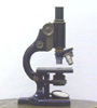
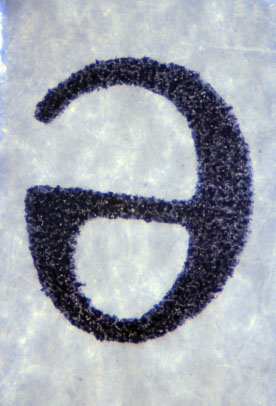
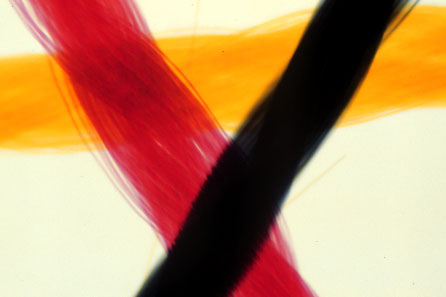


Microscope Lab
 

Title: Microscope Lab

Purpose:

  • to review how to use a microscope
  • to observe, draw, and describe human tissue cells

Materials:

  • prepared microscope slides
  • letter e
  • crossed threads
  • natural fibers
  • cork
  • cheek cells
  • blood smear
  • bone marrow smear
  • 3 types of muscle cells
  • 3 types of epithelial cells

Procedure:

  • Observe, draw, and describe what you see in all slides under low (40 x) and high (100 x) power
  • Label drawings
  • Label cell membrane, cytoplasm, and nucleus as appropriate

Results:

  • Drawings and descriptions

Discussion:

  1. Summarize what you did and comment on variety of cell types, shapes, and arrangements

Conclusion:

  1. State a conclusion about the variety of cell type, shape, or arrangement

Reflection:

  • (A personal statement about the activity, whether it relates to "real life," whether you liked it, suggested improvements.....)
letter e

threads

http://www.und.nodak.edu/dept/jcarmich/101lab/lab1/lab1.html

fibers
cork
cheek cells
blood smear
bone marrow smear

http://www.kumc.edu/instruction/medicine/anatomy/histoweb/cytology/cytology.htm

striated muscle
smooth muscle
cardiac muscle

simple squamous
epithelium


http://www.kumc.edu/instruction/medicine/anatomy/histoweb/epithel/epithel.htm

cuboidal epithelium


http://www.kumc.edu/instruction/medine/anatomy/histoweb/epithel/epithel.htm

BACK TO TOP

columnar epitlelium
stratified epithelium
pseudostratified epithelium
adipose tissue
Microscopy Links

Science and Photography through a microscope
http://www.pbrc.hawaii.edu/~kunkel/

Cell Cams
http://www.cellsalive.net/cam0.htm

see cancer and bacterial cells divide!

NanoWorld
http://www.uq.oz.au/nanoworld/images_1.html

Microscopy Primer
http://micro.magnet.fsu.edu/primer/index.html

Virtual Microscopes
http://micro.magnet.fsu.edu/primer/virtual/virtual.html

Eukaryotic Cell Organelles....a Shockwave Tutorial
http://www.ucalgary.ca/~doerksen/cell.html

Microscope Image Galleries: An Annotated List
http://mindquest.net/biology/cell-biology/misc/cell-pictures.html

Microscape: A Virtual Microscope with Images from Atoms to Ants
http://www.msa.microscopy.com/MicroScape/VL/Jawssmall.html

Microscopy: Size and Scale
http://mse.mcmaster.ca/research/micro/microsco.htm

Bugscope Gallery
http://bugscope.beckman.uiuc.edu/gallery/index.htm

not anatomy, but a great image gallery!

Microbe Zoo
http://commtechlab.msu.edu/sites/dlc-me/

Microscope images of the microbial world

BACK TO TOP

Animated Graphics Courtesy of
Jo's World